| Using Icon-NMR software to automate 1H NMR experiment on the
Bruker 400 MHz instrument. Experimental Chemistry II, CH 463/H |
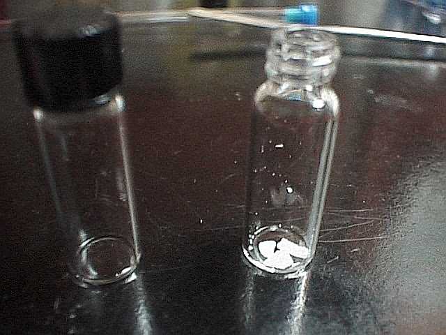
| Weigh or dispense about 10 - 20 mg of sample into a 4/5 dram
vial. |
 |
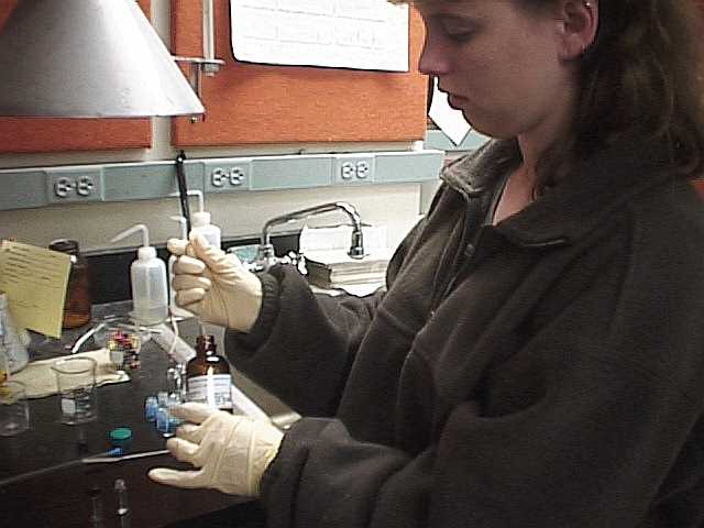 |
 Measure about 3/4 mL of solvent using a
disposable pipet. It should look like about this much. Measure about 3/4 mL of solvent using a
disposable pipet. It should look like about this much. |
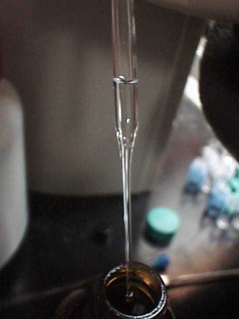
| Pipette solvent into vial and draw mixture back and forth until
a solution is made. |
 |
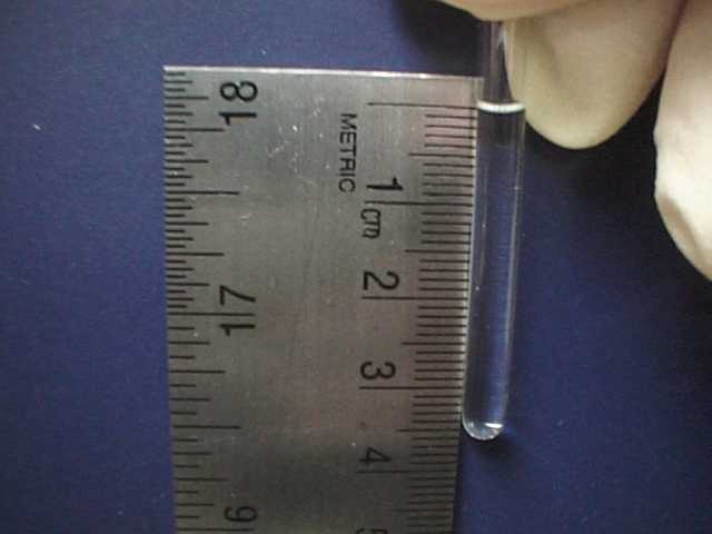 |
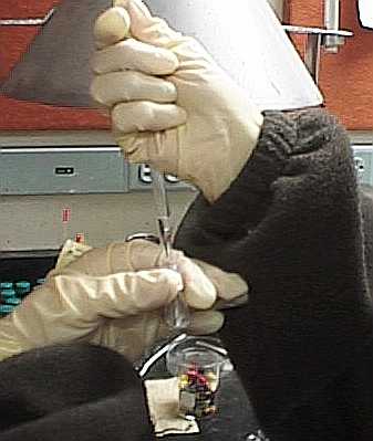
Dispense solution using pipette into the NMR tube. The
depth of solution should be about 3 cm. |
| The tube is then fitted with the spinner, and tube and spinner
are placed into the depth gauge. This puts the spinner at the correct height. |
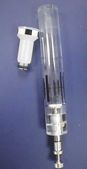 Spinner (left) and depth gauge (right) Spinner (left) and depth gauge (right) |
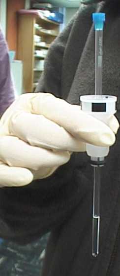 After depth gauging it, the tube should look like this
fitted with the spinner After depth gauging it, the tube should look like this
fitted with the spinner |
|
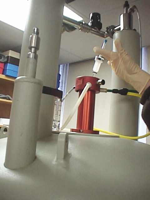
| Carefully put the sample tube in the probe chamber on top of
the magnetic |
 |
 |
The sample should look like this in the chamber. |
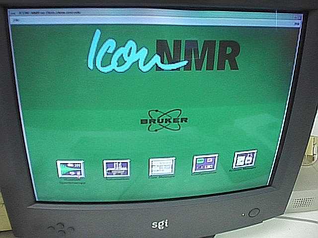
| Load ICON-NMR on the computer console for the Bruker 400 FTNMR
system in Gilbert 228. Consult the directions for using ICON-NMR (separate handout.) |
 |
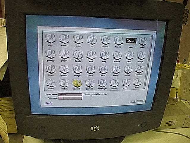 |
Login. |
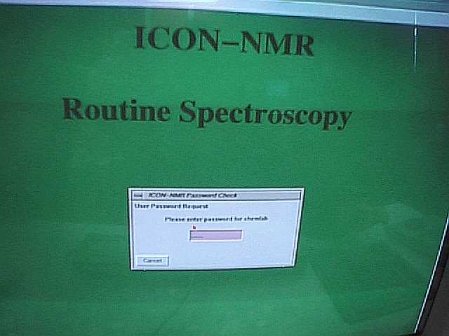
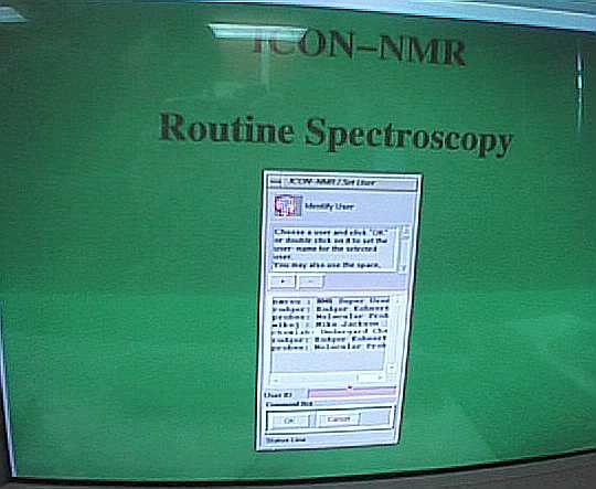
| Log in again to ICON-NMR Routine Spectroscopy |
 |
 |
|
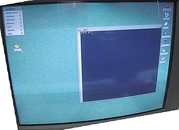
| Main program menu screen looks like this. |
 |
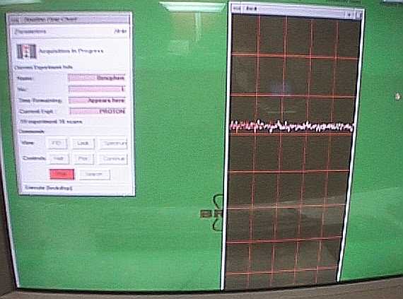 |
The program will automatically shim the sample. |
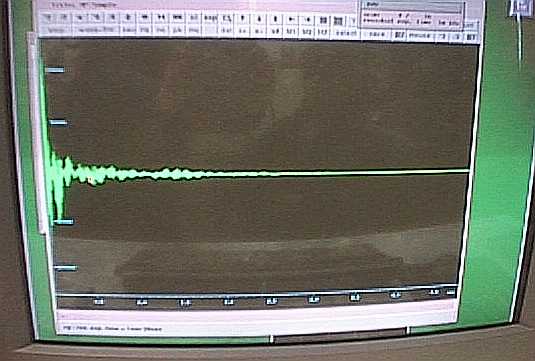
| Once shimmed the FID will be collected |
 |
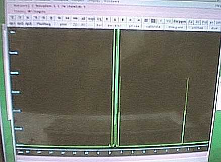 |
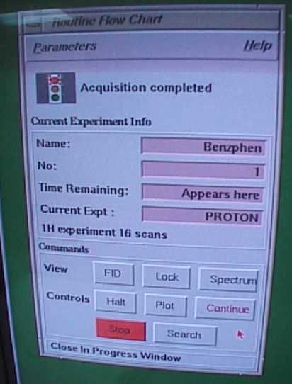
The FID will be transformed, phased and integrated for you
and a hardcopy will be printed. |
| To run another sample, select Continue |
 |