AFM Images from the VEECO/Digital Instruments Site
Hard disk
MFM image
Magnetic bits written with an MFM probe on perpendicular Co-Cr
media with a NiFe sublayer. The bits are about 180nm in size spaced 370nm,
giving an equivalent area density of ~5 Gbits/in2. 2.3µm scan courtesy Michael
Azarian, Censtor Corporation
Polymer blend
|
|
|
|
|
|
|
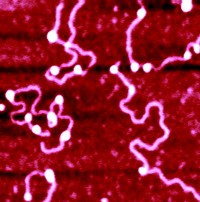
TappingMode image of nucleosomal DNA was the
highlight of the "Practical Course on Atomic Force Microscopy in
Biology," held at the Biozentrum in Basel, Switzerland, July 1998. Image
courtesy of Y. Lyubchenko.
Reconstituted
nucleosome cores were loaded onto plasmid DNA sequences (courtesy of Prof. Lohr).
The nucleosome is 11 nm x 5.5 nm and contains a protein core about which is
wrapped 1.75 turns of DNA. The horizontal streaks in the image are due to
incorrect "flattening" of the image during processing